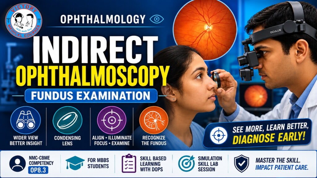
This competency-based simulation skills course is designed to equip undergraduate medical students with the knowledge, technical skills, and clinical approach required to perform indirect ophthalmoscopy for systematic fundus examination.
Indirect ophthalmoscopy is an essential ophthalmic examination technique that provides a wide-field, binocular and stereoscopic view of the retina, allowing examination of both the posterior pole and peripheral retina. The skill is particularly important for recognizing clinically significant retinal abnormalities and developing a structured approach to fundus examination.
The course uses a simulation-based DOAP (Demonstration–Observation–Assistance–Performance) methodology, allowing students to learn and practise the procedure in a controlled environment before applying the skill in clinical settings.
Learning Objectives
By the end of the course, the student should be able to:
- Explain the purpose and clinical importance of indirect ophthalmoscopy.
- Identify the components of the binocular indirect ophthalmoscope and condensing lens.
- Explain the basic optical principle involved in indirect ophthalmoscopy.
- Prepare the equipment appropriately for examination.
- Explain the procedure to the patient and obtain appropriate consent.
- Position the patient and examiner correctly.
- Hold and position the condensing lens appropriately.
- Achieve correct optical alignment.
- Obtain and maintain a clear retinal image.
- Perform a systematic examination of the optic disc, retinal vessels, macula and posterior pole.
- Demonstrate the approach to peripheral retinal examination.
- Recognize the appearance of a normal fundus.
- Identify important abnormal fundus findings.
- Recognize findings suggestive of conditions such as diabetic retinopathy, hypertensive retinopathy, papilledema, retinal tear and retinal detachment.
- Communicate significant findings appropriately and understand when ophthalmic evaluation is required.
- Demonstrate the skill safely and systematically in a simulated environment.
Course Content
Module 1 — Introduction to Indirect Ophthalmoscopy
Students are introduced to the purpose, indications and clinical importance of indirect ophthalmoscopy, including its advantages in examining the peripheral retina.
Module 2 — Equipment and Preparation
Students learn to identify and prepare the binocular indirect ophthalmoscope, condensing lens and other equipment required for the examination.
Module 3 — Optical Principle
The basic optical pathway is explained, including the role of the condensing lens in forming an aerial image of the fundus.
Module 4 — Patient and Examiner Positioning
Students learn appropriate patient positioning, examiner posture, headset adjustment and working distance.
Module 5 — Obtaining the Retinal Image
Students practise alignment of the examiner, ophthalmoscope, condensing lens, patient’s pupil and retina to obtain a clear retinal image.
Module 6 — Systematic Fundus Examination
Students are trained to follow a consistent examination sequence:
Optic Disc → Retinal Vessels → Macula → Posterior Pole → Peripheral Retina
Module 7 — Peripheral Retina
Students learn the principles of examining the peripheral retina using appropriate patient gaze and controlled examiner movements.
Module 8 — Normal and Abnormal Fundus
Students review representative fundus findings and learn to distinguish normal anatomy from important pathological changes.
Module 9 — Common Errors and Troubleshooting
Students identify common beginner errors, including poor alignment, incorrect working distance, excessive movement and incomplete retinal examination.
Module 10 — Competency Demonstration
Students independently perform the procedure in a simulated environment and undergo structured DOPS (Direct Observation of Procedural Skills) assessment.