Course Introduction
KOH Examination of Skin Scrapings for Fungus is a competency-based practical learning module designed for undergraduate MBBS students. The course introduces learners to the clinical and laboratory approach to the direct microscopic examination of skin scrapings for fungal elements using a potassium hydroxide preparation.
Superficial fungal infections are frequently encountered in clinical practice. Recognizing a suspicious lesion is important, but students must also understand how to obtain an appropriate specimen, prepare the KOH mount correctly, examine it systematically under the microscope, recognize fungal structures, and interpret the findings in conjunction with the clinical presentation.
This course uses a simulation-based, step-by-step teaching approach to connect clinical examination with practical laboratory diagnosis.
Learning Objectives
By the end of this course, the student should be able to:
Knowledge
- Explain the principle of KOH examination.
- Describe the indications for KOH examination of skin scrapings.
- Recognize common clinical features suggestive of superficial fungal infection.
- Identify the appropriate site for collecting skin scrapings.
- Explain the role of KOH in clearing keratin and cellular debris.
- Describe common fungal structures that may be observed microscopically.
- Explain the significance and limitations of positive and negative KOH findings.
Practical Skills
Students will learn to:
- Prepare the workstation appropriately.
- Follow hand hygiene and PPE requirements.
- Select an appropriate lesion and sampling site.
- Demonstrate safe collection of superficial skin scrapings.
- Label the specimen correctly.
- Prepare a KOH mount according to the institutional laboratory SOP.
- Apply the coverslip correctly.
- Allow adequate clearing.
- Perform systematic microscopic examination.
- Recognize fungal elements such as hyphae, yeast and pseudohyphae.
- Interpret and document the findings appropriately.
Clinical and Professional Skills
Students will also develop the ability to:
- Explain the procedure appropriately to a patient.
- Obtain consent when applicable.
- Maintain patient privacy and dignity.
- Follow laboratory safety precautions.
- Handle KOH safely.
- Dispose of specimens and contaminated materials appropriately.
- Correlate microscopy findings with the clinical presentation.
Course Content
Module 1 — Clinical Introduction
Students are introduced to superficial fungal infections and the clinical situations in which KOH examination may be useful.
The module demonstrates characteristic clinical appearances such as:
- Tinea corporis
- Tinea cruris
- Tinea pedis
- Cutaneous candidiasis
- Other superficial fungal infections
Students learn that clinical suspicion is the starting point for appropriate specimen collection.
Module 2 — Recognizing the Appropriate Lesion
Students learn how to examine a suspicious lesion and identify the appropriate area for sampling.
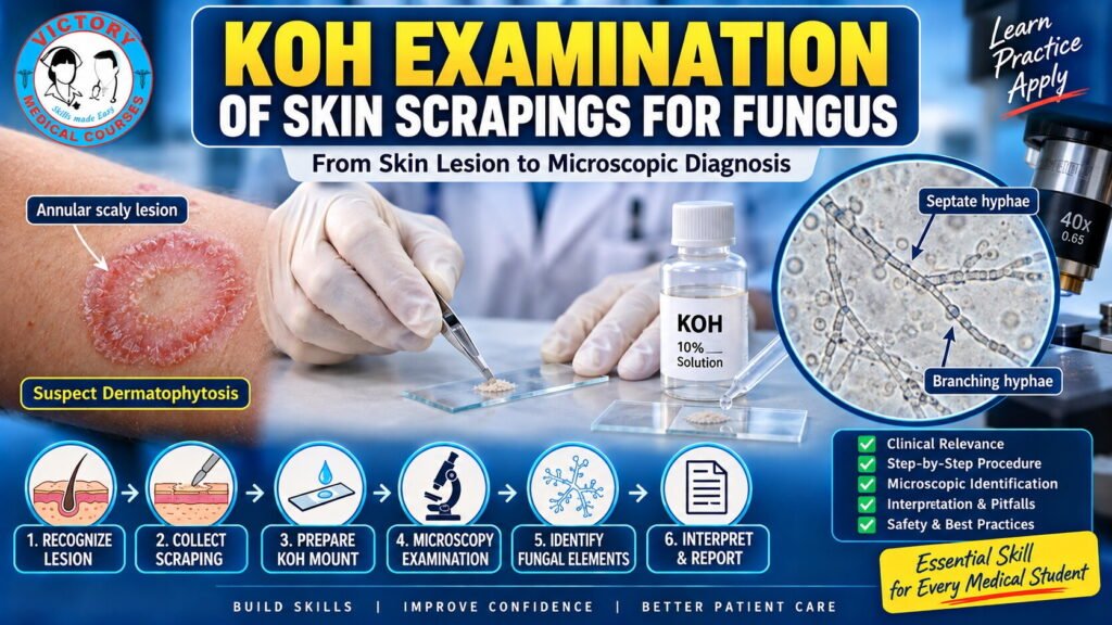
Particular emphasis is placed on the active scaly peripheral margin of a typical dermatophyte lesion.
The course emphasizes the principle:
Correct specimen selection is essential for obtaining a useful diagnostic result.
Module 3 — Collection of Skin Scrapings
Students observe and practice the appropriate technique for obtaining superficial skin scales.
The module covers:
- Patient/specimen identification
- Explanation of the procedure
- Hand hygiene
- PPE
- Appropriate sampling site
- Safe superficial scraping
- Avoiding unnecessary trauma
- Specimen transfer
- Slide labelling
- Safe handling of materials
Module 4 — Principle of KOH Examination
Students learn why potassium hydroxide is used.
KOH helps clear keratinous material and cellular debris, making fungal structures easier to visualize microscopically.
The fundamental concept is presented as:
Skin scraping → KOH preparation → Clearing → Microscopy → Identification → Interpretation
Module 5 — Preparation of the KOH Mount
The complete preparation process is demonstrated step by step.
Students learn:
- Placement of specimen on the slide
- Addition of KOH preparation
- Gentle distribution of the specimen
- Coverslip application
- Minimizing air bubbles
- Adequate clearing
- Preparation according to laboratory SOP
Students are reminded that KOH concentration, volume, clearing time and other technical parameters should follow the validated institutional laboratory protocol.
Module 6 — Microscopic Examination
Students learn systematic examination of the KOH preparation using a light microscope.
The module demonstrates:
- Initial low-power examination
- Locating suitable areas
- Higher-power examination
- Systematic scanning
- Recognition of fungal structures
- Avoidance of common microscopic artefacts
Module 7 — Recognition of Fungal Elements
Students are introduced to the appearance of common fungal structures.
Dermatophytes
Students learn to recognize:
- Branching hyphae
- Septate hyphae
- Hyphal fragments
Candida
Students learn about:
- Budding yeast
- Pseudohyphae
The emphasis is on recognizing fungal morphology rather than attempting species identification from a basic KOH preparation.
Module 8 — Interpretation and Clinical Correlation
Students learn how to interpret:
Positive KOH examination
Fungal elements are seen.
Negative KOH examination
No fungal elements are seen.
The course emphasizes that:
A negative KOH examination does not automatically exclude fungal infection.
Possible causes of a negative result include:
- Inadequate specimen
- Incorrect sampling site
- Insufficient material
- Prior antifungal treatment
- Technical limitations
- Observer limitations
Students therefore learn to interpret KOH findings together with the clinical presentation.