OTOSCOPY — EXAMINATION OF THE EAR
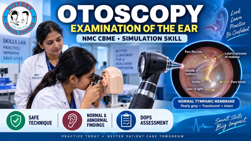
Otoscopy is an essential clinical examination skill for undergraduate medical students. This NMC CBME-aligned course provides a structured, simulation-based approach to examining the external auditory canal and tympanic membrane using an otoscope.
The course combines interactive learning, faculty demonstration, simulation practice, image-based interpretation, and competency assessment to help students develop confidence and accuracy in performing otoscopy.
Students will learn not only how to use an otoscope, but also how to prepare and position the patient, safely examine the ear canal, visualize the tympanic membrane, identify important anatomical landmarks, recognize common normal and abnormal findings, and document findings systematically.
COURSE OBJECTIVES
By the end of this course, students will be able to:
Knowledge
- Explain the basic anatomy relevant to otoscopic examination.
- Describe the components and appropriate use of an otoscope.
- Describe the normal appearance of the external auditory canal and tympanic membrane.
- Identify important tympanic membrane landmarks.
- Recognize common abnormal otoscopic findings.
Practical Skills
- Prepare the patient appropriately for ear examination.
- Obtain appropriate explanation and consent.
- Position the patient correctly.
- Select an appropriate ear speculum.
- Hold and manipulate the otoscope safely.
- Correctly align the adult external auditory canal.
- Examine the external auditory canal systematically.
- Visualize and assess the tympanic membrane.
- Identify the malleus, umbo, pars tensa, pars flaccida and cone of light.
- Assess tympanic membrane mobility when appropriate.
- Compare findings between both ears.
- Document findings using a structured approach.
- Represent important findings diagrammatically.
Professional Skills
- Maintain patient comfort and dignity.
- Follow appropriate infection-prevention practices.
- Recognize situations where examination should be stopped or modified.
- Communicate findings clearly and professionally.
COURSE CONTENT
Module 1 — Introduction to Otoscopy
Students are introduced to the purpose and clinical importance of otoscopy.
Topics include:
- What is otoscopy?
- Why is otoscopy performed?
- What structures can be examined?
- Importance of systematic examination
- Role of otoscopy in undergraduate clinical practice
Module 2 — Otoscope and Equipment
Students learn to identify and prepare the equipment required for examination.
Topics include:
- Otoscope
- Handle and light source
- Viewing head
- Magnifying lens
- Ear specula
- Speculum selection
- Equipment checks
- Infection prevention and safe handling
Module 3 — Patient Preparation and Positioning
Students learn the correct approach to the patient before beginning the examination.
Topics include:
- Patient identification
- Introduction
- Explanation of the procedure
- Consent
- Patient comfort
- Appropriate positioning
- Head stabilization
- Communication during examination
Module 4 — Correct Otoscope Technique
This module focuses on the practical technique of performing otoscopy.
Students practice:
- Correct otoscope grip
- Stabilization of the examining hand
- Pinna manipulation
- Adult ear canal alignment
- Gentle speculum insertion
- Maintaining visualization
- Avoiding unnecessary force or trauma
Module 5 — Examination of the External Auditory Canal
Students learn to systematically assess the ear canal for:
- Patency
- Cerumen
- Erythema
- Edema
- Narrowing
- Discharge
- Foreign body
- Skin abnormalities
Emphasis is placed on safe examination without blind instrumentation.
Module 6 — Examination of the Tympanic Membrane
Students learn to identify and describe the normal tympanic membrane.
Key features include:
- Colour
- Translucency
- Integrity
- Position
- Surface appearance
- Pars tensa
- Pars flaccida
- Handle of malleus
- Lateral process
- Umbo
- Cone of light
Module 7 — Tympanic Membrane Mobility
Where appropriate and equipment is available, students are introduced to pneumatic otoscopy.
They learn the basic interpretation of:
- Normal mobility
- Reduced mobility
- Absent mobility
The course emphasizes appropriate patient selection, safe technique and correlation with other clinical findings.
Module 8 — Normal and Abnormal Otoscopic Findings
Students are exposed to representative otoscopic images and learn to recognize and describe common findings.
Examples include:
- Normal tympanic membrane
- Cerumen obstruction
- Otitis externa
- Acute otitis media
- Tympanic membrane perforation
- Retraction
- Myringosclerosis / tympanosclerosis
- Other visible abnormalities
The emphasis is on recognition and accurate description, rather than expecting undergraduate students to make specialist-level diagnoses from an isolated image.
Module 9 — Documentation and Clinical Communication
Students learn how to communicate their findings in a structured manner.
Documentation includes:
- Side examined
- External auditory canal findings
- Tympanic membrane colour
- Position
- Integrity
- Landmarks
- Mobility when appropriate
- Abnormal findings
- Comparison between ears
- Diagrammatic representation
Students are encouraged to replace vague descriptions such as “ear normal” with a structured clinical description.
Module 10 — Competency Assessment
Students demonstrate the complete procedure through DOPS/OSCE-based assessment.
Assessment includes:
- Hand hygiene
- Patient identification
- Explanation and consent
- Positioning
- Equipment preparation
- Speculum selection
- Otoscope handling
- Pinna manipulation
- Safe insertion
- Canal examination
- Tympanic membrane examination
- Landmark identification
- Recognition of findings
- Documentation
- Patient safety and communication